
In most cases, tooth root caries occurs in elderly patients over 60 years of age. This category occupies about 60-90% of all cases of revealing a disease at the dentist's appointment.
According to the widely used classification, depending on the affected part of the tooth, the following types of caries are distinguished:
- tooth decay of the tooth;
- cervical caries;
- tooth decay;
- radical caries.
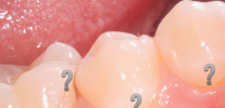
What is the difference between tooth root caries from cervical and radical? Tooth decay is located deep beneath the gum, violating the integrity of the tissues of the bare and invisible root during a visual examination.
Cervical carious cavities develop only near the gingival margin on the buccal as well as on the labial surfaces of the enamel. That is, these are areas visible to the eye.
The photo below shows a comparison of cervical caries with root caries:


As for basal caries - it is formed along the exposed roots of the teeth on the lingual, contact and buccal surfaces. This means that in this case, we can also see with the eye the areas of tooth decay.
To summarize the preliminary result. Tooth decay is tooth decay, not visible to the eye, but can lead to serious problems. Now consider the important nuances in more detail.
The main causes of tooth root caries
Tooth decay under the gum most often develops in people due to serious gum diseases: atrophy, dystrophic processes in the tissues or as complications after their treatment. Otherwise, tooth decay is called "cement decay." Typically, destruction begins with the cervical zone on the open surface of the root (see photo).

It is interesting
Microorganisms and products of their vital activity penetrate the root cement and wash out the mineral components from it. In this case, organic components (collagen) are preserved. Over time, the active activity of microflora under the gum leads to the destruction of a thinned cement layer that does not have a reliable mineral base. Is developing hidden carious process at the root of the tooth.
Let us consider in more detail the main causes of root caries:
- Often, root caries develops with the formation of a gingival pocket and the accumulation of food debris in it. The gingival pocket is a consequence of the violation of the normal attachment of the gums to the cervical part of the tooth, when the gum can literally “move away” from the basal zone. In this case, tartar and plaque can most often be found there. The activity of microorganisms that feed on carbohydrate residues of plaque makes it easy to form a carious process on an uncoated root with the subsequent formation of a cavity.
- In addition, root caries may be due to complications of cervical caries. That is, the destruction of enamel visible with the eye gradually deepens inward: from the neck of the tooth to its root.
- Also, the cause of tooth root caries can be a delayed treatment of major caries, when not only the tooth crown and cervical part are destroyed, but it also comes to the root itself.

- And finally, root caries can occur when the crown is placed poorly on the tooth or after its expiration date. In this case, a gap is formed between the edge of the crown and the gum, from which the tooth peeks. It is in this area that plaque actively accumulates and over time a stone forms. Destruction of the tooth at this point can even lead to breaking off the crown of the tooth.

Characteristic clinical manifestations
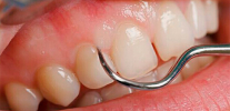
The main feature of tooth root caries is the absence of complaints in most cases in the early stages of the onset of decay. Hidden carious lesion under the gum For a long time, it may not be able to detect itself at all, but after the “exposure of the gums” or the formation of a large carious cavity, the following symptoms appear:
- Pain from thermal stimuli (cold, hot), chemical stimuli (mainly from sweets) and mechanical (when solid food gets under the gums).
- Aesthetic disturbances visible to the eye. This is due to an increase in the lesion area and the unification of caries at the root base with cervical defects. The following photo shows an example:
- Discomfort when eating. This is often caused by tooth extensions and impaired support from the ligamentous apparatus that holds it in the socket. In this case, mobility develops with the appearance of unpleasant sensations associated with chewing food and getting it into the cavity of the root caries.

On a note
Since in the early stages of destruction it is practically impossible to feel caries at the root base, you should consult a dentist for the purpose of diagnosis once every 6 months, especially if there are serious age-related changes in the gums, as well as with painful phenomena in them with the above symptomatology.
Features of revealing hidden caries
To successfully detect root caries, the dentist uses a number of techniques.
For example, sensing using a sharp probe is widely used. A mandatory requirement for this technique is a preliminary removal of stone and plaque from all surfaces of the teeth, since hidden foci are found precisely under the thickness of the dental plaque.
An acute probe is a dental instrument safe for healthy tooth tissues for examining and diagnosing hidden foci of tooth decay. When examining a probe with a probe, the doctor carefully inserts it into hidden foci to detect enamel roughness, chips, medium and large defects.
 is widely used to diagnose hidden foci of tooth decay.")
In addition, radiological research methods are often used to diagnose root caries. They are ideal for the initial forms of root caries, even when there is no carious cavity. In this case, most often used:
- Bite-wing-roentgenogram;
- Parallel radiography method;
- Orthopantomogram.
The photo shows an example of an x-ray with a clearly visible pathology at the root of the tooth:

It's important to know
It is necessary to fulfill all the recommendations of the dentist on the diagnosis of hidden foci, since the time spent on a long search for root destruction will pay off with timely started tooth treatment and the prevention of serious complications, often leading to its loss.
Modern methods of root caries treatment
Depending on the location of caries, its area, depth, severity of the process, the general situation in the oral cavity, etc., the dentist chooses the optimal tactics, which consists of the general principles of treatment of tooth root caries. These include:
- Professional oral hygiene. This is an important step in achieving the necessary results, since most often only the elimination of dental deposits allows you to access the carious cavity, treat it in the cleanest conditions without an additional infected focus.
- Elimination of plaque delay factors on artificial crowns and exposed roots: correction of fillings overhanging the gingival margin, replacement of bad dentures, elimination of dentoalveolar anomalies (for example, crowding of teeth that interferes with normal oral hygiene by brushes and pastes).
- Treatment of initial and superficial caries of the root base without filling. Most dentists have a wide arsenal of fluoride-containing preparations (varnishes and gels) with or without an antiseptic.

It is interesting
Well-proven drugs that contain 0.05-2% sodium fluoride, aminofluoride, 4% titanium fluoride with 1-5% chlorhexidine or triclosan, 0.4% tin fluoride.Deep fluoridation involves the use of a dentin-sealing liquid that contains fluoride crystals and copper ions in the diagnosis of tooth root caries. It is advisable to use fluorides in combination with calcium preparations (10% calcium gluconate solution and 0.5-1% sodium fluoride solution for applications on the root lesion site).

It should also be noted the treatment of superficial and deep caries with a filling technique. When treating subgingival cavities, a difficult situation often arises: the inability to isolate a place for a future filling well from saliva, gingival fluid, blood, etc.
As a result, the doctor carefully protects the gum from unnecessary damage: it can carry out diathermocoagulation (removing excess overgrown gums with a tip heated to high temperatures), retract (correct overhanging edges) of the gums with special threads soaked in a hemostatic solution. The whole procedure is carried out, of course, with good anesthesia to achieve optimal end results for the treatment of root caries.

Treatment of the cavity is carried out under water cooling of the tooth, an oval cavity cleared of caries and infection is formed more often with additional areas for better fixation of the future fillings. It is treated with antiseptics and sealed.
Currently, the complexity of choosing a material for filling is associated with different types of cavity location, its shape, the presence of various interfering factors: saliva, blood, gingival fluid, etc. It is difficult to put the material “dry”, and modern composite (“light”) fillings are very sensitive to the humid environment.
Acceptable seal materials currently:
- Amalgams. Unfortunately for dentists, this material is less and less used because of the complexity of organizing the mixing of the material. These are the most durable fillings that are mixed with mercury, and therefore require the creation of conditions for the protection of personnel.
- Compomers. These are materials that combine the positive properties of composites and glass ionomer cements. However, there are times when the properties of the composites in them interfere with successfully “catching” the seal from the compomer for guaranteed fixation on the tooth.
- Glass ionomer cements. This is the best option for sealing deep subgingival defects, as this material is best glued in a humid environment. Also, fluorides are introduced into it to restore the normal mineral structure of the tooth over a long period of time.

Prosthetics for deep defects
In addition to the treatment with traditional filling techniques, the methods of prosthetics of the cavities with tabs and crowns are also used. A tab is, in fact, an artificial filling made of metal or ceramic, which is made by a dental technician, and the orthopedic dentist fixes cement or special adhesives into a prepared and cleaned carious cavity.
The convenience is that the tab replaces large cavities with messages with the subgingival part, and the risks of fixation are minimal. Since the tab has a stump - “tail”, which is fixed in the root of the tooth, it reliably remains cemented and performs its functions in full.
A crown is an artificial cap made of metal or a combination of metal and ceramics, which is securely fixed to the tooth and covers all its surfaces from the effects of infection from the oral cavity.
The picture below shows an example of restoration of a decayed tooth using the tab and crown:

A correctly placed insert or crown provides excellent protection of the tooth from possible complications of root caries: fracture, fracture of the crown, “decay”, various gingival disorders. However, it is worth remembering that before setting the crown, the tooth is either filled according to the usual technology, or the tab is fixed on it, and then the crown. Only this gives a positive result for the long term.
Clinic pricing
Based on the criteria of the modern pricing policy of most clinics, it is useful to keep in mind the following points.
- For any material there are certain indications. A good doctor will never do the tooth job that is contraindicated in this clinical situation. If it is enough to put an inexpensive filling made of glass ionomer cement, the doctor will fix it, and if the tooth is damaged so much that you need an insert or seal + crown, the dentist will draw up a treatment plan, explain the cost and save the tooth in full.
- Filling materials are always cheaper than materials for prosthetics. Seals are much cheaper than aesthetic and functional crowns or inlays. However, there are exceptions.


Timely treatment of tooth root caries is an important stage in preserving the tooth, preventing serious complications from the tissues surrounding the tooth, adjacent teeth, and the bite as a whole. In the presence of the slightest signs of abnormalities on the part of the gum or suspicion of a carious cavity hidden under the gum, you should immediately contact a professional dentist for help.
In the normal condition of the oral cavity, it is enough to visit the dentist's office once every 6 months for the purpose of a routine examination for early detection of caries. Take care of your teeth and be healthy!
Interesting video: complications of root treatment
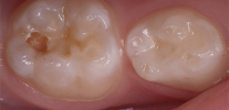
An example of the treatment of deep caries
Root apex resection
Generally speaking, there is no such nosology as hidden caries in medical reference books and encyclopedias. Although the dentists themselves and their patients by this term ...
From the point of view of the causes and development of the pathological process, caries under the gum is no different from caries on tooth surfaces visible to the eye ...
Most people have wondered more than once about what the word "caries" means, even despite a more or less clear understanding of the processes, ...
On my upper teeth, as I understood from your article, cervical caries, how is it treated? And is it possible to do something without tearing it apart? (while there is no discomfort, but I'm afraid of the consequences).
There is a complex of treatment: special caries grinding and installation of photographic fillings. Visiting a doctor 2 times a year. To undergo prophylaxis: polishing, applying cleansing gel and fluoridation. Brush your teeth after eating. Healthy teeth!
On my lower front teeth, the gums are moving away from the teeth, the deposits have been removed, but the gums are also moving away. What can be done to strengthen? I'm afraid I'll lose my teeth. FRONT, this is a disaster. And I'm afraid that tooth decay will appear on the roots.
Hello Olga! I think that you should consult a periodontist. This is a specialist in the treatment of diseases of the gums and oral mucosa. The treatment plan will be drawn up on the basis of a thorough clinical examination. Almost always, the process is a set of measures aimed at restoring the periodontal attachment. For example, you may need gum repair, periodontal dressings for gums, curettage of periodontal pockets, etc. The choice of treatment tactics belongs to the doctor after a full-time examination. Health to you!
I have four front lower teeth grind off ... Can I restore them? The trip to the dental technician did nothing.
Hello! It seems to me that you are confusing a dental technician with an orthopedic dentist ("prosthetist"). I think that you went to the orthopedist, who did not offer you anything suitable.If we are talking about tooth abrasion, then restoring such teeth is extremely difficult. The fact is that your bite is most likely convenient for you, so increasing the height of your teeth will become a traumatic option for your bite. It seems to me that the reasons for the orthopedic dentist were quite there to refuse you. I am sure that you should turn to another orthopedist, who has the ability to gradually “raise” the bite with the help of special caps that will have to be worn for medical purposes. So the bite will gradually form to such an extent that something can be done with an increase in the height of the front lower teeth. Most often, this is done already with crowns at the last stage of treatment. However, this approach takes on average about 6-12 months: it all depends on the required millimeters of height.
Hello, I have caries of the front tooth, with the edge of two larger ones ... In general, the gum above this tooth is darker and slightly swollen. I think that caries is also under the gum. Will it be deleted for me, or can I be saved?
Hello! The final verdict according to your description will be made only by the dentist, putting you in a chair. It all depends on the depth, area of the lesion, tooth mobility, its position, bite and other nuances determining the fate of the future. I can immediately say that most often the front tooth can be saved. Often, conservation is complicated by technical nuances: treatment of the canal, inflammatory processes at the root, subsequent restoration of the tooth with or without a pin, etc. An alternative or more reliable method of preserving a tooth may be to create a tab on it + crowns. Before that, the doctor reliably obturates the canal and prepares the tooth already for orthopedic work. In general, each doctor may have an opinion on the possibility of preserving a particular tooth with its features of carious lesions, etc. For my part, I advise: having received the verdict "tooth extraction", I recommend that you go through another 1-2 consultations in other clinics in order to have some kind of objectivity in your clinical situation. Have a good diagnosis and treatment!
Hello! I have stones on my four lower teeth. At one of the teeth, half of the root fell out, at the same tooth root caries began, and after that cervical caries began. This tooth has become very weak, even when hit with a soft test, it starts to hurt terribly. I am very afraid of losing it. Please tell me, can doctors help this tooth if it starts to cause pain when it gets very soft food? What will happen if a dental instrument hits? A tooth can fall out altogether.
Hello! In fact, pain from irritants while eating is not a problem. The tooth can always be pulped and sensitivity will disappear. However, if you have periodontitis, it is unlikely that the doctor will take up not the most “strong” lower front tooth, although all this is decided individually. The least information at the moment is about tooth mobility: I can’t evaluate either its “staggering” or the degree of “exposure” of its cervical region and the extension of the clinical crown. All this is evaluated by the dentist and, as a result, makes a verdict. Such teeth are saved according to the situation (the need for it as a whole in relation to the bite, other teeth, etc.). The approach in this case is complex: it begins with the treatment of the canal and its filling and ends with work with adjacent teeth and splinting them together. Neighboring teeth most often also have to be treated.
Hello. I was diagnosed with tooth decay. No treatment was offered. The tooth did not hurt on its own. Only when the doctor pounded him with an instrument. They suggested removing it to avoid complications. The tooth has been removed, but I do not see caries at the root, as in the photo. I see gutta percha sticking out of the root.Could it be that the tooth was removed in vain? Or perhaps caries inside a temporary filling?
Hello! I think that your suspicions are not in vain about the extraction of a tooth that has no root caries, but the complication after the treatment is the withdrawal of gutta-percha from the top of the root, and also, possibly, the perforation of the tooth bottom.
It seems to me that the situation was as follows: the dentist decided to treat this tooth, but encountered some obstacles. He could not overcome them with the help of further conservative tactics, and the only correct way out in this situation to prevent your future torment was to remove the tooth.
You write about a temporary filling and gutta-percha, which indicates that the tooth was treated intracanal. I think that it was definitely not a question of root caries, but there was something serious. Perhaps the colleagues did not want to substitute their own colleagues, or there was something else, but what difference does it make now if the very fact of the removal is completed, and it will be difficult to prove anything now. The tooth you have in your hands could be sent for examination, because the removed gutta-percha outside the root is already a complication that the first doctor who treated your canals made it. As for the rest, this is only after removing the temporary dressing and assessing the condition of the bottom of the extracted tooth. Well, the picture would not hurt at that moment when the tooth was still in its place in the dentition - as a justification for the fact that there were clear indications for removal.
Hello, I had an X-ray of a 6 tooth (3-6), on which root caries was found. The doctor drilled a tooth and laid the medicine to kill a nerve. The tooth ached, the gum turned gray and moved away from the tooth. After 2 days (since it was a weekend), I again went to the dentist. She said that this medicine passed through the cavity in the tooth and because part of the root was exposed, it healed it (I did not see the cavity, information from her words). After that, she cut off part of the gum, saying that she would recover better, and laid the medicine. At the next visit, she tried to clean the roots, but after spending 5-7 minutes (the others did not even look), she said that the roots were thin and she did not see them, and in general it was not necessary to open them. Laid down a “mummifying” ointment (in her words, it will dry out the nerves remaining in the roots) and put a seal, saying that if it hurts, it must be removed.
A week has passed, the tooth hurts when biting. I don’t want to delete it. Can you comment on the failure of root treatment? Maybe because of them the tooth continues to hurt? And can this lead to root inflammation?
Hello! I think that this is a routine method of treatment. Most likely, there are certain problems in this institution. Mummifying method is not the best option for saving a tooth. During treatment, there was also a serious mistake that led to a complication. I will not blame the doctor - I do not know in what conditions he has to work.
The tooth hurts when biting with a high degree of probability due to the aggression of the mummifying paste introduced into the beginning of the unpassed channels. There is also a risk that the tooth hurts due to an infectious process.
It’s difficult for me to comment on the refusal of canal treatment, I can’t know if the canals are objectively so complex, or is this all the result of the clinic’s routine approach to treatment. This treatment may well lead to inflammation. In some cases, these teeth can become seriously ill after 1-3-5-10 years. It all depends on how well they are mummified. Yes, this old methodology also has rules, without violating which it is quite possible to get some success with all the disadvantages that have forced many countries to abandon this drug and the method of preserving teeth.
My advice is this: treat according to a modern technique, before it’s too late, a tooth from a highly qualified doctor in a clinic where there is good equipment and time to work normally, possibly on your complex canal systems. In principle, this is quite real, and the question of price is purely individual, and is discussed with the dentist in advance.
In my upper row of teeth, caries of the anterior tooth root. The doctor of the municipal dentistry refused to treat - said, delete. And I feel sorry for the tooth, what to do, help!
Hello! A public institution often refuses complex teeth for various reasons: there is no funding, motivation, desire, materials, skills, experience in this direction, etc. That is why I recommend contacting a private clinic or several clinics in order to have a more complete picture of the possibilities of preserving a tooth, as well as the prices of various options. Often, during tooth decay, a tooth is pulped out and a crown is placed for reliability. There is also another way to save: gum retraction and caries treatment with glass ionomer cements or (less commonly) composites. It all depends on what your clinical situation is and the condition of the adjacent teeth, taking into account the bite. In general, a lot of things affect, therefore, an examination by an experienced specialist is necessary. So I recommend getting advice in different clinics, since in more than 50% of cases, consultations are positioned as free or inexpensive.
Hello! I got the canals in the lower seven under VHI insurance. The picture clearly shows that the filling material came out in a thin stream below the root itself in 2 channels. The jaw below does not ache much and gives in the ear. Above is a temporary seal. On the neck of this tooth is an open hole from cleaning caries. Temporary filling does not hold.
What to do if the tooth has not calmed down, and how will the seal stay on the neck of the tooth?
Hello! The fact that the material is taken outside the root is a complication. Whether it will result in something serious for the future, even if the symptoms disappear in a few weeks, is not known (in order to at least roughly predict, you need to know what material is derived). Authoritative dentists oppose the removal of materials beyond the apex of the root.
As for the possibility of holding the filling in the cervical region - of course, there are some nuances, but usually the material of choice is glass ionomer cement or light-cured filling. When performing the technology of working with this area, the seal holds for years without any problems.
Hello! After the CT scan, I was diagnosed with tooth decay of the root of the 7th tooth. Four months earlier, I had an 8 tooth removed (complicated, traumatic extraction), and then I also had a CT scan and with 7 tooth everything was in order. Is there a root caries, the doctor’s fault? I have to remove other teeth, I don’t know whether to look for another one, since the doctor himself was very pleased.
Hello! For 4 months, root caries would hardly have formed, but not the fact that at present it generally has a place to be. I could comment in more detail if I had pictures in my hands. If you trust the doctor - trust completely, otherwise check according to the image data. At the same time, keep in mind that in the dentist's chair it is much easier to understand the clinical situation than in absentia (that is, you need to consult another doctor for an in-person consultation, and it’s better to go to another clinic).
If by doctor’s fault you mean some complication during the removal of the 8th tooth, which entailed caries of the root on the 7th tooth, then this is impossible.If this gap between the teeth in the presence of a wisdom tooth was constantly “clogged” with food, then the root caries on the 7th tooth developed over the years, and after the extraction of the end tooth it simply became clearly visible (especially if the cavity is deep). From the picture, even a small carious process in the gum zone can be detected. Maybe this is just your case.
So here either trust the doctor, or check it. Choose you.
Hello, I turned to a free clinic the other day, they removed the carious root, and the crown itself has long been destroyed. And this root began to bother at night. And now I understand that it could possibly have been saved in a paid clinic. They also plan to remove the wisdom tooth and send it for x-ray. You know, I don’t want to lose my teeth at 26 years old. What do you advise to do? And what should I put in place of a live gum? I read about bridges and about implantation, it seemed to me a better way out. I don’t want to lose two more teeth because of the bridge. So scary is all ...
Hello! Implantation would be the best option, so as not to process adjacent teeth under the bridge crowns. However, there are contraindications for implantation, which can become a serious obstacle to implant engraftment in the bone, so that the optimal prosthetics options in each case are selected individually.
As for the need to remove a wisdom tooth, it is impossible to answer this question without additional information, since a lot depends on the condition of the tooth (whether it is retinned or semi-refractory, as it is located in the jaw bone, does it injure the mucous membrane of the cheek, are there any inflammatory processes on the roots, etc.) .d.) I recommend finding a good doctor in a paid clinic (ask friends, for example) and make a joint decision with him.