
Generally speaking, there is no such nosology as hidden caries in medical reference books and encyclopedias. Although the dentists themselves and their patients actively use this term.
The point here is that hidden caries is the common name for any caries that cannot be found on teeth without special diagnostic methods. This is its main difference from caries in other manifestations: teeth look like absolutely healthy, but the pathological process in them can be so developed that treatment of the disease requires not only the removal of part of the dentin, but also depulpation (“nerve removal”).
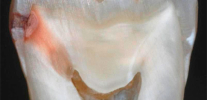
The photo below shows the hidden caries in the extracted tooth (located on the contact surface, that is, in the place of contact with the adjacent tooth). The affected area reached the pulp chamber:

In many cases, latent caries accompanies the ordinary and plural, and only in exceptional cases does it develop independently among other healthy teeth. Such cases are most rarely diagnosed and most often lead to very deep tooth damage.
Tooth damage in case of hidden caries
In general, damage to teeth in the form of a cavity with hidden caries differs little from those for cases distinguishable to the naked eye. The nature and extent of such damage is determined by the age of the carious process and its localization on the tooth. So, hidden caries can have the following localizations:
- On the proximal (contiguous) and distal (posterior) walls of the tooth. Here, its manifestations on enamel are very difficult to notice with a mirror or probe.
- Under improperly placed fillings or crowns. If bacteria from the oral cavity can enter the space between the tooth tissues and the filling material, the carious process is likely to begin to develop here.
- In the areas of the tooth located under the gum (food particles often accumulate here and are not cleaned with a toothbrush).
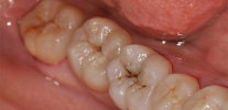
- In fissures, natural fossa of the “back teeth”. If the damage to the enamel is small, then it is not always possible to notice it with a mirror and a probe, and therefore even with the actual presence of external manifestations, such caries can be classified as hidden.
In the photo - typical fissure caries with minimal external manifestations:

Damage to a tooth with hidden caries is similar to that with a visually diagnosed form of the disease. However, the specificity of the process leads to the fact that latent caries is usually found only in the middle and late stages of development, when it covers a significant part of the dentin and peri-pulp tissue. Accordingly, when an affected tooth is opened, extensive damaged cavities are detected.
The photos below show an example of hidden caries in a tooth under a filling:


And in this picture there is a roentgenogram of a tooth with hidden caries at a rather late stage of development. It can be seen that the area affected by caries came close to the canal with the nerve:

From the practice of the dentist
A typical clinical case: in a patient, among two teeth affected by caries, there is one, seemingly healthy, without visible damage. Logic suggests that if two teeth are affected, it is likely to be affected by caries and located between them. I assign a radiograph to the patient in order to check the integrity of the internal tissues. A well-developed caries near the posterior wall of the tooth is seen on the radiograph. The defeat had not yet reached the pulp, but penetrated deep into the dentin. In this situation, it was enough to clean the damaged area and seal.
Clinical manifestations
Hidden caries practically does not manifest itself. Since there is either no visible damage to the tooth enamel with it, or they are small, until the pulp is involved in the pathological process, the patient usually does not experience discomfort when hot or cold liquids get on the tooth, chews it normally.
In rare cases, the affected area expands towards adjacent areas of enamel and with a closer look at the tooth you can notice darkish spots. The photo below shows an example of such a hidden caries under an old seal:

When the pathology spreads to the innervated areas of the tooth, the patient begins to experience pain when exposed to cold or hot teeth, as well as acidic, sweet or salty foods. The first few weeks or months, this pain is not very strong and does not always occur. Gradually, it intensifies and reaches a stage at which the patient is forced to go to the clinic. In such situations, depulpation (“nerve removal”) is almost inevitable.
What is the danger of hidden caries?
The main danger of latent caries is the inconspicuous development of the pathological process to such stages that require either depulpation or tooth extraction in general.

In addition, with the development of caries on proximal surfaces, it is possible to “transition” to the adjacent tooth, and sometimes with the development of the disease in it in a hidden form.
On a note
This requires clarification: for some reason, many people are sure that caries can be “transmitted” and infect an adjacent tooth, but this is not so, caries is a local process and not “contagious”. It is important to understand that in the literal sense of the word, there will be no transition to another tooth. Perhaps there is something else: if latent caries developed on one tooth with the formation of a cavity, then self-cleaning of the gap leaves much to be desired. A constant accumulation of carbohydrate food debris causes an aggravation of the process in the first tooth and the beginning of a new destruction in the neighboring one.

In rare cases, either without pain, or with a patient’s unwillingness to see a doctor, the carious area can cover most of the space filled with dentin. This situation is fraught with the fact that the tooth, in fact, turns into an empty shell, which can crumble even with a small load.
The photo below shows hidden caries with a large lesion area (x-ray), a significant part of the tooth crown is covered by pathology:

Diagnosis of the disease
Modern diagnostic methods allow revealing latent caries at any of its localization and at any stage of development. But in order to get to the use of these methods, the dentist must have suspicions of a carious process, even if indirect. Often in the absence of patient complaints and when examining the oral cavity, latent caries remains unnoticed and develops safely until pain in the affected tooth appears.
In many cases, latent pathology develops in parallel with carious lesions, which are clearly visible on other teeth. In this case, an experienced dentist correctly assesses the general cariogenic situation in the oral cavity, understands that with frequent and multiple lesions of other teeth, the likelihood of developing hidden caries is very high, and prescribes diagnosis using special methods. These include, for example:
- An x-ray in which areas with hidden caries look darker than healthy neighbors.
- Transillumination and luminescent diagnostics. Both of these methods reveal hidden carious tooth damage at any stage.
- Laser diagnostics.


It is almost impossible to independently detect hidden caries. Before the development of pulpitis, it will not be detected when tapping the tooth and treating it with cold or hot water.
Therefore for early diagnosis of hidden caries regular preventive visits to the dentist are very important.
How is latent caries treated?
The treatment of latent caries in most cases consists in the removal of the affected cavities and further filling of the tooth. Moreover, if the pathology develops in the cervical zone or under the gum, the dentist has to adjust the soft tissues surrounding the tooth.
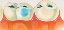
The pictures below show examples of preparation of cavities:


In some cases, latent caries are detected at the stage of stain or surface damage to the enamel without penetration into the dentin. In these cases, for successful treatment, it is possible to do without an autopsy. The doctor can strengthen the enamel with calcium and fluoride preparations to restore it (remineralization).
In cases where a hidden carious process is found under a shaky filling, the damaged areas are cleaned and a new filling is installed in their place.
Such treatment is usually painless and only occasionally can cause tingling. If sharp pain occurs in the patient during dentin stripping, this indicates involvement of the pulp in the process and requires removal of the nerve with further filling of the canals.


In most cases, surgical treatment of latent caries is carried out using anesthesia (local anesthesia), even if “drilling” is not required deeply. Usually this question is consistent with the patient before treatment, and the decision is made mutually.
In rare cases, with a large-scale damage to the tooth, excision of all tissues damaged by caries, up to the level of the gums, with the subsequent installation of a crown, or even tooth extraction and implant implantation, is required. As a rule, with regular visits to the doctor and constant examinations, it is possible to avoid the disease at such neglected stages and to preserve the teeth that have been filled, but native.
Prevention of hidden caries does not differ from that of its other forms: it is necessary to observe oral hygiene, remove food debris stuck between the teeth, limit the use of sweets in between meals, and use calcium and fluoride preparations when prescribed by a doctor. And most importantly - regularly undergo examinations at the dentist. Hidden caries is a good example of the fact that a visit to the doctor is necessary even without pain in the teeth and other obvious signs of the development of the pathological process.
Causes of caries and effective methods of protection against it

And so, in fact, the preparation of the cavity and installation of the filling takes place live
By the term "internal caries", an ordinary patient in a dental clinic usually understands a disease that affects tissues deep under the tooth enamel. ...
The decompensated form of caries is, in fact, an intensively developing caries, a pathological process that is very active in solid ...
The simplicity or, conversely, the complexity of the diagnosis of caries directly depends on its location on the teeth and stage of development. Consequently, from the stage, ...
Thanks for the article, a fully disclosed topic. As a patient, I was looking for a way to clarify secondary and latent caries. This is an x-ray. Thank you)) Good luck!
My doctor did not notice hidden caries at a routine examination. After 6 months, the tooth began to crumble, and it turned out that the caries was already such that you need to put a crown ... That's how I lost my tooth. And by the way, the doctor is very professional and expensive, from a private clinic.
But the doctor told me that there was no hidden caries on the X-ray + the nerve was removed in that tooth, and said that it could ache the gums.But more than a week has passed, and the little tooth still hurts from below and between. What to do in this case?
Hello! The situation is difficult for remote search for the cause of pain. It can be anything: from a doctor’s mistake in diagnosis to banal carelessness. I guess one of the reasons:
1. Poorly treated tooth canals;
2. Hidden caries on an adjacent “living” tooth;
3. An improperly formed contact point (the gap between the teeth), due to which the gum is injured.
You can send your tooth picture to the site’s mail - I will look and comment on it. With the help of images, the amount of information on a clinical case is often significantly increased. Be that as it may, your situation requires more careful consideration to exclude periodontitis in a tooth already treated in the canals, as well as possible pulpitis (or caries) in one of the neighboring ones.
Hello, a tooth with nerves hurts and gives down to the jaw. There was a filling, 5 years ago there was caries, treated, after a week the tooth ached, the doctor said that everything will pass - the pain went away. But over time, she began to get sick again, went to the doctor - he sent for an X-ray, said that everything was fine and he could not bother, but replaced the seal (in my opinion, there was caries under it). But the tooth still hurts, and now the pain has intensified. What could it be? Thanks for the answer.
Hello, Tatyana! The symptoms described by you indicate a possible inflammation of the pulp of the tooth (pulpitis). And despite the fact that on the x-ray, as the doctor told you, “everything is fine”, you need to understand the causes of the problem, not expecting that everything will go away by itself. Constant pain in the tooth indicates the possible presence of inflammation, which, if nothing is done, may well lead to tooth loss. I recommend contacting an experienced dentist-therapist to clarify the situation (without examination, via the Internet in your case there is no way to figure out the true causes of pain).
I had it. It’s a jamb when sealing and cleaning the canals. Inflammation at the end of the root is not treated, the tooth is removed.
These schools are treated endodontically for the most part. Should consult with other specialists before removal. Perhaps in your case there were good chances to save a tooth. Sorry.
Hello. About 2 years ago I treated a tooth. Sealed. Now a dark spot has appeared around the seal. What does it mean? That they badly brushed a tooth before filling, did not completely remove the caries? Need to go to the doctor again? So I do not want to ((
Hello, Cyril. Still, it is necessary to consult a doctor - apparently, there was a violation of the marginal fit of the seal to the tooth. The reasons can be very different, it is not at all necessary that the doctor “badly brushed his tooth”. A period of 2 years is sufficient to cause such a problem. It is important to quickly replace the seal, since otherwise the situation will only worsen and can lead to complications (for pulpitis, for example).