
The term “bite” in orthodontics refers to the type of closure of both jaws with each other in a static (habitual, effortless) position. The so-called orthognathic bite is recognized by the doctors as the most physiological - if it is available, a person can fully perform the functions of chewing, breathing and speech, and therefore, live a full life.
On a note
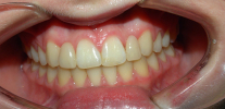
The orthognathic bite is most consistent with a smooth and beautiful “Hollywood smile”, however, as we will see below, some deviations from the ideal may be present even in this case.
Let's see what kind of bite it is, how it is formed, what are its main signs, and whether orthodontic treatment may be required with an orthognathic bite ...
Why is an orthognathic bite so important for dental health?
An orthognathic bite creates optimal conditions for the functioning of the entire dentition. The teeth occupy natural, normal places for them in the rows, without obstructing hygiene with a toothbrush, and close together in a position most suitable for effective chewing of food.

With such a physiological bite, an adult normally has no significant gaps between the teeth, which means that there are no additional conditions for the accumulation of food debris. When food in significant quantities regularly remains in the interdental spaces, decay processes begin, which entails halitosis (bad breath), gum disease, demineralization and softening of the enamel of the lateral walls of the teeth.
On a note
It is known that any deviations from normal occlusion, whether crowded teeth, their abnormal position, the presence of three (large gaps) are a risk factor for tooth decay, since often the incorrect position of the teeth contributes to the accumulation of food particles and does not allow to completely clean the enamel with a brush from plaque and bacterial film.
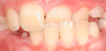
The photo below shows how malocclusion can impede dental hygiene:

With an orthognathic bite, periodontal tissues are not subjected to excessive stress and stress, while when the teeth deviate from the normal position, foci of tension in the gums and ligaments are often created, the natural process of nutrient exchange is disrupted, which can cause a decrease in the adjacent part of the gums and exposure of root cement a tooth.
In the presence of occlusion pathologies, patients may notice the appearance or exacerbation of the so-called wedge-shaped defects, increased sensitivity to acidic, cold, excessive tooth erasure.

In addition, many orthodontists, based on extensive clinical experience, believe that in the presence of even the slightest deviation from the orthognathic bite, the temporomandibular joint (TMJ) reacts with a kind of ligamentous apparatus remodeling. Crunching, clicking when opening the mouth and chewing, pain and muscle tension, regular headaches, sometimes not stopped even by taking analgesics, are typical signs of TMJ malfunctioning due to deviations from the correct bite.
On a note
Among the possible problems due to malocclusion, bruxism is worth mentioning separately - excessive compression and grinding of teeth, usually at night.
An interesting fact: many general practitioners (pediatricians, therapists) often believe that helminthiasis, that is, helminthic invasion, is the cause of bruxism. Doctors believe that the presence of helminths in the body causes hunger in a person, which causes him to excrete large amounts of saliva in his sleep and involuntarily make chewing movements.However, there is no scientific evidence for this hypothesis ... While among the scientifically proven causes of bruxism, dentists distinguish stress and the above described TMJ disorders.
With an orthognathic bite, as a rule, diction is not disturbed, and the smile looks beautiful.
Signs of orthognathic bite
The orthognathic bite is characterized by a number of specific signs - let's look at them in more detail in order to have a clear idea of what a “Hollywood smile” is.
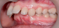
In the language of orthodontists, an orthognathic bite is the closure of dentition according to Engle's I class, namely: the mesial-buccal tubercle of the first molar of the upper jaw is located in the inter-tubercle space (fissure) of the first molar of the lower jaw. Thus, the so-called occlusion key is formed.
An example is shown in the figure below:

Occlusion is determined by the nature of the displacement of the lower jaw relative to the upper, in which a particular number of teeth come into contact with each other. This is an important concept for dentists, thanks to which you can understand the cause of various complaints of patients.
There are four main types of occlusion: anterior, right lateral, left lateral and, finally, central occlusion. Central occlusion (central bite) is the position of the lower jaw relative to the upper, in which the maximum number of teeth are in simultaneous contact with each other.
So, central occlusion with an orthognathic bite is characterized by several signs:
- Dental sign - with central occlusion, the chewing surfaces of the lateral teeth and the cutting edges of the front teeth are in close contact with each other, each tooth of the upper jaw is closed with two teeth of the lower jaw (except for the last molar of the upper jaw and the first incisor of the lower jaw). The upper incisors overlap the lower ones by one third of the height of their crown, the first molars close according to Engle's I class, the middle lines passing between the central incisors of the upper and lower jaws are in the same plane;
- Muscular sign - the muscles of the lower jaw should be in a state of myodynamic equilibrium (that is, the correct closure of the teeth occurs naturally and does not require efforts from a person);
- The joint sign - the head of the joint and the capsule should be at the beginning of the protrusion of the joint - tubercle.

On a note
Anterior occlusion, in turn, is characterized by the presence of contacts only in the area of the frontal group of teeth. With lateral occlusions, the side to which the lower jaw moves is called the working, and the opposite side - the balancing one.
In 1972, six occlusion keys were first described, which orthodontists use to this day. They were derived on the basis of a study of 120 plaster models of the jaws of people with an orthognathic bite, and in honor of the author, these keys are called Andrews keys:
- The first key coincides with the definition of an orthognathic bite according to Engle;
- The second key describes what the normal value should be the inclination of the tooth crowns along the entire length of the dentition;
- The third key describes the degree of inclination of the teeth lingually;
- The fourth key says that normally teeth should be positioned in an arc without tilting or turning along the axis, that is, they should be standing exactly;
- The fifth key indicates the absence of gaps between the teeth;
- The last (sixth) key suggests that the occlusal surfaces of the chewing teeth should not be in the same plane, but three-dimensionally, thereby forming occlusal curves (the Spee curve and the Wilson curve). Dentists use these curves for treatment planning and proper tooth placement.

Interesting fact
Patients under normal conditions close their mouth in the position of their usual occlusion, and this position does not always correspond to central occlusion.Meanwhile, this position is important in orthodontics and orthopedic dentistry for prosthetics and staging, so doctors often have to resort to various tricks to determine the central ratio of teeth.
What other types of bite are the physiological norm
In addition to orthognathic, there are other types of bite that allow you to fully chew food, talk and maintain oral hygiene at a normal level:
- Direct bite - with it, the ratio of molars corresponds to the first class of Engle, however, the incisors are joined together by joint. A significant drawback of a direct bite is that this type of closure with age leads to abrasion of the cutting edges of the incisors;

- Biprognathic bite - characterized by a normal ratio of teeth in the lateral parts, however, the incisors of the upper and lower jaws are excessively extended forward and are closed by cutting edges;
- A prognathic bite is another type of physiological bite in which the anatomical alveolar bone and maxillary incisors are tilted forward toward the upper lip;
- Progenic bite (“pro” - forward, “genus” - chin) - in the anterior part of the dentition there is a reverse incisal overlap, that is, the chin is advanced and the lower incisors overlap the upper ones (mesial occlusion).


")
Generally speaking, these types of occlusion do not require compulsory correction, but sometimes patients do not like their appearance, and then the orthodontist, without violating the ratio of molars, resorts to treatment, trying to change only the position of the frontal group of teeth.
Periods of physiological bite formation: what is important for parents to know
The formation of the dentition of the child is usually divided into several periods. Even if there are genetic prerequisites for the formation of an orthognathic bite, it is important that each of the stages proceed smoothly, without serious pathologies. Let's analyze each of the periods and see what should be paid attention to the parents of the baby.
The first is the period of neonatality and the onset of teething of temporary teeth. In this period, the sucking reflex dominates, and thanks to the function of sucking, the development and growth of the jaws, especially the lower jaw, occurs.
At this time, gum semicircular ridges without teeth are visible in the baby’s mouth. The lower jaw is in a distal position with respect to the upper jaw, that is, behind it - doctors call this phenomenon infant retrogenia. The structures of the temporomandibular joint are not yet expressed, which allows the baby to carry out active sucking movements.

At the age of 6-7 months, the eruption of the first temporary teeth begins, they erupt in a certain order (usually the lower teeth appear first, then the upper ones).
The table below shows the standard procedure and terms of teething (in parentheses are the serial numbers of teeth adopted in dentistry):
| Temporary tooth | Duration of teething, months |
| Central incisors (I) | 6-7 |
| Side incisors (II) | 8-12 |
| Fangs (III) | 12-16 |
| The first temporary molars (IV) | 16-20 |
| Second Temporary Molars (V) | 20-20 |
Deviations of 2-3 months in one direction or another from the dates indicated in the table are considered normal, however, if the teeth have not erupted during this time, this is an occasion to visit a pediatric dentist to find out the cause of the delay.
It is interesting
Sometimes a baby is born already with natal teeth in the oral cavity. This is not a reason for panic, but is only an individual feature of the development of the baby.
So, the bite period of temporary teeth follows. The formation of such a bite ends by 3-3.5 years. This stage is characterized by the following features:
- The surfaces of the lateral teeth are located in a vertical plane;
- In the area of the posterior teeth there are tight contacts, the front upper incisors overlap the lower ones.
- The teeth are arranged in a row without three (spaces).
An intermediate period of preparation for a change of teeth is also distinguished. This stage is characterized by the presence of three - milk teeth diverge, preparing a place for permanent ones. The growth of the jaw bones is observed from front to back.
At this time, the chewing function dominates, and the lower jaw actively grows, moves forward, the cutting surfaces of the incisors undergo a process of physiological erasure:

Then comes the period of change of temporary teeth to permanent ones - it begins with the eruption of the permanent first molars.
The table below shows the sequence and time of eruption of permanent teeth:
| Permanent teeth | Teething time |
| First molars (6) | 6-7 years old |
| Central incisors (1) | 7-8 years old |
| Side incisors (2) | 8-9 years old |
| Premolars (4) | 9-11 years old |
| Fangs (3) | 10-12 years old |
| Second Premolars (5) | 11-12 years old |
| Second molars (7) | 12-13 years old |
Usually in this period, parents notice the presence of crowding of the teeth in the baby, especially the lower incisors. This compensatory phenomenon is caused by the fact that permanent teeth are larger in size and require more space for themselves. The location also plays a role - for example, the lower lateral incisors are more lingual, preparing a place for massive permanent fangs.
The lower front incisors can bend lingually or rotate along the axis, that is, stand slightly sideways. Some crowding can be seen in the upper central incisors.
Orthodontists call this stage the “ugly duckling” stage, but, nevertheless, this is a normal stage of bite formation. After the eruption of fangs, the incisors are aligned, and the trem between the teeth disappear by themselves.

On a note
If there is a risk of a child swallowing a tooth due to its premature mobility, then such a tooth must be removed (there is also a risk of airway obstruction). After the removal of such teeth, careful monitoring of the orthodontist is required in order to create conditions for the further normal development of the baby’s dentofacial system. It is necessary to "keep" a place in a row so that it is not occupied by "neighbors" and a permanent one could erupt in the place of the tooth that has fallen out. To do this, the doctor can make removable plate apparatus with an artificial tooth.
Is an orthognathic bite necessary?
Even with an orthognathic bite, it is still sometimes possible to detect abnormalities, in some cases requiring orthodontic treatment.
The most common occlusion abnormalities include:
- The mismatch of the size of the teeth and jaws, which entails the development of the crowded position of the teeth. Clinical experience shows that anatomically molars are quite massive and require more space for themselves - thus, when teething, they put pressure on the entire dentition, and the incisors turn around on their axis, taking up less free space;
- Teething outside its normal position - may be due to improper laying of the tooth germ in embryogenesis, or early loss of milk teeth;
- Tremas, as well as diastema (a crest between the first incisors of the upper jaw). After the appearance of permanent fangs in the dentition, the diastema normally closes independently, but if this does not happen, the dentist or orthodontist can refer the child to the surgeon to correct the frenulum of the lip. Sometimes a diastema appears as a result of the presence of a supernumerary tooth in the upper jaw in the area between the central incisors, which can be detected only by x-ray. As for the three, they appear compensatory, if the teeth are smaller than they should be for the existing size of the jaws;
- Retention of milk teeth in a row - this phenomenon is caused either by the absence of the rudiments of permanent teeth, or by the incorrect position of the rudiment itself in the bone, which prevents it from erupting.

In all these cases, despite the general physiological nature of the bite, a doctor's intervention may be required.
Modern approaches to the treatment of malocclusion of the first class
With a lack of space in the dentition and slight deviations in the position of individual teeth, sometimes the most correct tactics will simply not intervene, because how treatment in this case can aggravate the situation, be long and tiring for the patient.
If there is a shortage of space more than 4 millimeters, the patient may be offered to make transparent caps with the rearrangement of individual teeth in a more correct position. In such cases, on the gypsum models of the patient's jaws, the doctor who carefully removes the teeth that need to be moved and relocates to another position - then, according to this model, the mouthpiece is made of transparent material.
 allow you to effectively correct bite without resorting to the use of bracket systems.")
With more complex malocclusion, the doctor may recommend treatment with a bracket system so as not to upset the first-class ratio. In this case, a large expansion is not necessary, therefore, ligature bracket systems are considered more acceptable for this purpose. It is the ligature braces that allow the doctor to clearly monitor the inclinations of individual teeth, and the arches in these systems are narrower, which guarantees the absence of excessive expansion of the dentition.
As you can see, even with an orthognathic bite, the help of an orthodontist is sometimes out of place. Be healthy!
What is a right and wrong bite: comments of an orthodontist
What is important for parents to know about the formation of the correct bite in a child
Permanent bite is the relationship of the dentition of permanent teeth with the complete closure of the upper and lower jaws. Speaking simply, t ...
In simple terms, the distal bite is such an anomaly of the bite, in which the teeth of the upper jaw are strongly advanced forward about ...
Anomalies of the occlusion are various deviations from the normal arrangement of the dentition relative to each other. Similar deviations may ...