
Permanent bite is the relationship of the dentition of permanent teeth with the complete closure of the upper and lower jaws. In simpler terms, this is a fully formed bite, when all milk teeth are replaced by permanent ones.
Compared to a temporary bite, a permanent bite is characterized by a number of important features - further we will examine in detail the stages of its formation, interesting nuances of the transition from a milk bite to a permanent one, as well as modern methods of treating malocclusion and situations when such treatment is difficult ...
Important nuances of the transition from milk to mixed bite
Orthodontists pay special attention to the period of the late milk bite, when preparations are made for the replacement of temporary teeth with permanent ones. Already at this time, when examining the child's oral cavity for a number of signs (not always obvious), future problems in the position of the teeth can be suspected.

Normally, the shape of milk teeth coincides with the shape of teeth in a permanent bite, but the crowns of temporary teeth should be proportionally wider, especially in the area of temporary molars (i.e., chewing teeth with numbers 5 and 6). Wide crowns in this case prepare a place for two future permanent teeth at once - premolars.
In turn, temporary incisors (front teeth) have more convex outlines and are normally slightly inclined palatine, since the roots of the permanent teeth located in the bone exert pressure on their roots.
The size of the child’s teeth and dental arches is much smaller than during the period of a permanent bite. In children under 4 years old, the lower jaw occupies a posterior position, but when the period of active growth of the jaws and heads of the temporomandibular joint begins, the lower jaw shifts forward (to some extent this is due to the nature of the child’s nutrition - cessation of sucking and active chewing of food). With normal growth of the jaws between the temporary teeth of the child, gaps (three) appear - this indicates the correct development of the dentoalveolar system, and in no way should be considered as a pathology by parents (as it sometimes happens).

On a note
If an adult or teenager has gaps between the teeth, this is really a sure sign of a pathological bite, or diseases of the tissues of the oral cavity (for example, gingivitis or periodontitis).
The roots of milk teeth dissolve over time, and they fall out, giving way to new permanent teeth. But sometimes it happens that milk teeth remain in place, and despite the fact that the child is growing, the change of individual teeth does not occur.
This may be due to several reasons:
- The child may not have the germ of a permanent tooth in the bone. In practice, when parents are questioned, it is usually possible to find out that the family has such a pattern, and relatives may not have separate teeth or even groups of teeth. Obviously, in such cases, pathology is associated with heredity;
- Or, a permanent tooth cannot leave the bone due to its improper position or interference from neighboring teeth.
In the x-ray below, the permanent teeth in the bone, formed under the milk and ready to push them, are clearly visible:

In any case, only the dentist can understand the reason for the delay in changing temporary teeth after the examination - an x-ray will be taken. After evaluating the image, the orthodontist discusses treatment options.
For example, if an embryo is absent, then after fixing the bracket system, the lost milk tooth is retained until the orthodontist creates enough space in the row for further prosthetics of the desired tooth.
If there is a permanent tooth in the bone, but it has little room for eruption, or it is too deep, or lies in the wrong position, then after fixing the bracket system and creating a place for the desired tooth, the tooth is gradually “pulled out” by the surgeon, fixing it on it, first, the orthodontic button, and then tying it to the orthodontic arch.
Characteristics of a permanent bite and what factors can affect it
An important stage in the formation of a permanent bite begins long before the eruption of permanent teeth - even at the stage of mineralization of their primordia. Mineralization with the formation of full-fledged hard tissues occurs inside the gums, therefore, only a doctor can evaluate how this process is going, by conducting an X-ray examination of the child. Mineralization begins in the first months of the baby’s life, and even after the appearance of the tooth, this process does not end immediately, but continues for another 1-1.5 years for the complete “maturing” of the root of the permanent tooth.

The mixed bite period begins with the eruption of the first permanent tooth. There is a certain order and terms of teething of permanent teeth:
- as a rule, molars are the first to erupt - 6 teeth at the age of 5-6 years;
- between the ages of 6-8 years, incisors begin to erupt alternately (first the first incisors on the lower jaw, then on the upper);
- soon the lateral incisors of the lower and upper jaws appear.
Upper incisors are usually located with a slight lingual inclination, and they are larger than temporary teeth. Their appearance coincides with the period of growth of the upper jaw, and initially they are located in their place tightly and without gaps.
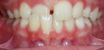
When the lateral incisors erupt, they exert pressure on the central upper teeth that have already appeared, due to which the central incisors diverge, forming a physiological diastema (crest) and leaning toward the lips. However, normally after the eruption of permanent fangs on the upper jaw, the diastema closes itself. Orthodontists often call this stage of development of the dentofacial system the “ugly duckling” stage, but after the teething of a child’s fangs, the teeth of both the lower and upper jaw are aligned.
After the incisors appear on both jaws, a period of physiological rest occurs, lasting 1-1.5 years.
Between the ages of 9 and 12, the second stage of teething of permanent teeth begins. At this time, the fangs change, premolars - “fours” and “fives” appear on the lower jaw (the eruption order is 3-4-5, and on the upper jaw, on the contrary, 4 tooth appears first, followed by a canine, and after that - 5 tooth).
The second last molars appear - the last but one - 7, and behind them the wisdom teeth (third molars, that is, 8s).
It is important to understand that although there are more or less certain periods of teething in a permanent bite, but they are relative, and in practice there may be some deviations from them that do not lead to serious consequences.
The figure below schematically shows the age characteristic of teething of certain teeth:

Thus, in a permanent bite, a person has 28-32 teeth, the shape of which resembles that of milk teeth. In addition to the main groups of teeth already in the temporary bite, 4 new ones appear - premolars.
We should also talk about the emerging “eights”, which often cause a lot of trouble due to the negative impact on the bite ...
On the teeth of wisdom and their possible impact on the bite
Since wisdom teeth are the last to appear in the oral cavity, when teething they can change the position of the remaining permanent teeth, pressing on their “neighbors” and displacing them, thus providing a place for themselves.

On a note
The nature of human nutrition since the time of primitive ancestors has changed significantly.The molars, especially the wisdom teeth, were needed by a person earlier for grinding hard bones and chewing hard raw meat. The load on the chewing apparatus was very significant, and from this the size of the teeth and jaw bones was larger. Today, people mainly eat soft, thermally processed foods, the chewing load has become smaller, so the size of the jaw bones has decreased. As a result, there is initially little space in the jaw for wisdom teeth.
Knowing this fact, many pediatric dentists and orthodontists recommend removing wisdom teeth before they begin to erupt, so that they do not ruin the teenager’s permanent bite in the future. Statistics of studies on this topic show that teething wisdom causes problems such as crowding, tooth rotation, displacement of the first molars and the formation of a pathological bite in 35-40% of cases!
However, the removal of the innocent and even not yet had time to erupt the "eights" - a moot point, and some experts do not approve of this practice. Indeed, for many people wisdom teeth do not lead to problems, regularly perform their chewing function and, moreover, in old age they come in handy when prosthetics are necessary.
Be that as it may, the decision to remove the wisdom teeth should be made by the orthodontist together with the patient after the examination. Even at the stage of drawing up a treatment plan, the doctor discusses the issue of removing the “eights” - it is often better to start from this stage, and after healing and recovery begin to align the bite.

On the other hand, it happens that already in the process of treatment the orthodontist, seeing the dynamics, recommends removing the wisdom teeth. There is a technique for removing the rudiments of still unformed wisdom teeth - this option is in many cases less traumatic than the removal of fully formed teeth, but requires a certain skill from the surgeon.
On a note
An unambiguous indication for the removal of wisdom teeth is their incorrect location in the jaw bone - when they are tilted towards the roots of adjacent permanent teeth or cheeks, because of which they cannot cut through, and often cause inflammation of the surrounding tissues.
How to understand that something is wrong with the G8? The process of their appearance, even in norm, can cause certain inconveniences: gums itch (especially at night), salivation increases, some adults complain of mild pain in the area where the tooth is cut. All these phenomena are a variant of the norm.
But there are alarming symptoms that should not be ignored:
- Persistent temperature increase above 37.5 degrees;
- Swelling and redness in the area of the cutting tooth;
- Severe pain, difficulty chewing and swallowing.
In all these cases, especially if the dynamics are negative and eventually only gets worse (pains are stronger, the temperature is higher), you do not need to wait - consult a doctor as soon as possible! In advanced cases, inflammation against the background of difficult teething wisdom can threaten the life of the patient.
Types of permanent bite: what is considered the norm, and what is a pathology?
It is customary to distinguish several types of physiological bite, that is, allowing to fully realize the chewing function:
- Orthognathic bite - is considered the most aesthetic and most favorable for maintaining a healthy state of soft tissues of the oral cavity and temporomandibular joints. The orthognathic occlusion has the following features: teeth in the lateral part are closed according to Engle's I class, namely, the anterior-buccal tubercle of the upper “six” is located in the inter-tubercle fossa of the lower 6th tooth. The front teeth of the upper jaw overlap the incisors of the lower jaw by no more than one third.All teeth on both jaws are in close contact with each other. Moreover, they have a certain slope, ensuring their smooth and correct position;

- Direct bite - in this case, the ratio of the lateral teeth is preserved according to the first class of Engle, and the teeth in the anterior section join butt-to-butt (over time, this can lead to their abrasion);
- Previously, it was also customary to attribute to physiological ones a progenic bite (pro - forward, genus - chin). That is, with this type of bite, the chin is pushed forward. In the anterior part of the dentition, a reverse incisal overlap is observed, that is, either the lower jaw is advanced, due to which an abnormal ratio of permanent molars is observed, or the teeth of the lower jaw are inclined towards the chin. However, at the same time, this position of the jaw bones can be called abnormal, corresponding to the mesial bite;
- Prognathic bite - in this case, the upper jaw is in front of the lower, the upper jaw incisors are tilted forward by their cutting edges, due to which the lower jaw incisors close at the posterior point, the lateral teeth abnormally close: the anterior-cheek tubercle of the first upper molar of the upper jaw is in front of the inter-tubercle fossa of the molar of the lower jaw. This ratio of the first molars can also be called abnormal - it corresponds to a distal bite;
- Biprognathic bite - both jaws are shifted forward relative to the base of the skull, in the anterior part the incisors are closed by cutting edges, and in order to achieve this closure, the incisors of the upper and lower jaws are tilted forward.


On a note
With progenic, prognathic and biprognatic bites, chewing function can be quite normal, although the bite is pathological and can lead to additional problems in the future - therefore, the orthodontist may suggest correcting it.
When evaluating a permanent occlusion in an adult patient, the orthodontist pays attention to how a person closes his teeth during a conversation, with the lower jaw moving sideways and forward. This closure of the teeth with various movements of the lower jaw relative to the upper is called occlusion.
Allocate front and side occlusions. An assessment of occlusion is important in understanding whether there is a malfunction in the normal chewing process. This allows you to anticipate early tooth erasure, muscle overstrain of the maxillofacial region, loosening and, in severe cases, tooth loss.
The photo below shows what the erasure of teeth leads to due to improper occlusion in the anterior region:

Normally, the lateral teeth of the upper and lower jaws should not remain in the same plane when closed. That is, the molars must have a certain inclination, so that with various movements of the lower jaw, the lateral teeth do not lose contact with each other. In the presence of the correct inclination of the teeth, conditionally drawn lines are formed - occlusal curves:

Given the shape of the occlusal curves, an orthodontist can reveal signs of an irregular permanent occlusion during a close examination of the patient's dentition.
The front teeth can also close incorrectly. For example, it is not uncommon for the incisors of the upper jaw to overlap the incisors of the lower jaw excessively - sometimes so much so that the lower teeth cut their edges into the palate, injuring it. Such an overlap in the anterior region is called a deep bite:

The reverse situation is when the teeth in the anterior part do not close at all, or their overlap is minimal. This bite is called open:

An open bite is often found in the side of the dentition:

Another type of irregular permanent bite is the cross, and the cross closure can be both in the anterior and in the lateral section. This anomaly occurs due to a mismatch in the size of the upper and lower jaws or their individual segments:

As a rule, a malocclusion combines pathological closure of the lateral and front teeth, so drawing up a treatment plan for a particular patient sometimes becomes a difficult task for a doctor.
Bite formation in adolescence
The adolescent period is a test for the entire body of the child, and the dentition is no exception. Under the influence of sex hormones, there is a natural surge in the growth of the body, after which the growth slows down and gradually stops.
Puberty in boys and girls is different:
- Girls begin to grow up about two years earlier than boys. The start of puberty in girls is the beginning of the formation of the breast and the appearance of the first pubic hair. The peak growth of girls is observed 1-1.5 years after the appearance of the first signs of puberty. After another year and a half, the final stage of maturation of the girl begins - the beginning of the menstrual cycle, after which growth slows down and ends after another year and a half;
- Boys, unlike girls, do not have such a clear separation of the changes that are taking place. It is known that active growth in boys begins about 3-3.5 years later than in girls, and also ends later. The first sign of the onset of puberty in boys is considered to be rapid weight gain. The growth spurt begins a year after weight gain - at this time the boy is losing weight, he has hair in the groin area. At the third stage of growing up - after 8-12 months, the growth of muscles and bones occurs, the shape of the body changes. Two years later, the last stage of growing up begins, growth stops, the first facial hair appears.
All these changes, one way or another, affect the formation of a permanent bite, and not always for the better.

Experienced orthodontists usually try to begin treatment of the bite precisely during the peak of the child’s growth, because the growth of the jaw bones as a whole coincides with the growth stages of the entire skeleton, and at the peak of growth, treatment is most effective and the result can be achieved faster.
On a note
To understand how a child is shaped and whether he has a reserve of growth, an orthodontist can direct him to an X-ray of his hand to see if the so-called growth zones on his arm have closed or not.
Thus, if there are problems with bite during teenage years, then you should not delay the visit to the orthodontist - it is better to start solving the problem during this period.
Bite correction in adult patients
Adult patients have their own specifics, and this may be associated not only with physiology. For example, adults want to have a beautiful smile, but, at the same time, often do not want others to see braces and any other devices on their teeth. Although in recent years there has been a fashion for braces - their presence is perceived not as a sign of imperfection, but as a sign of prosperity and prosperity.

To date, there are three areas of aesthetic orthodontic appliances that allow you to correct the anomalies of a permanent bite in adults.
The most popular option is vestibular braces (that is, glued to the teeth from the lips), made of materials similar in color to tooth enamel, which makes the bracket system inconspicuous.
Aesthetic vestibular braces are made of three types of materials:
- Plastic;
- Ceramics;
- Sapphire.
On a note
Braces made of plastic are the least expensive, but they are fragile, easily broken, have the property of being painted. So, for example, if you like tea, coffee, cigarettes, then these braces will quickly turn yellow.
Compared to metal braces, aesthetic braces to a greater extent require the doctor to fix accurately. Re-sticking or unfastening the locks during the treatment process can damage the grill, which ensures that the bracket itself adheres to the tooth, and it will not hold, which means that in any unusual situations, the patient will have to pay for a new bracket.
In addition, such systems are more expensive than metal counterparts by about 20-30 thousand rubles.For example, a set of “simple” metal braces that are tied to an arc with rubber bands, ligatures, costs an average of 20 thousand rubles, and the aesthetic option, depending on the material of manufacture, can cost 40-50 thousand rubles.
It should be noted that sapphire braces are the most durable and do not lose their aesthetic and functional properties, ceramic ones behave a little worse - they can be painted in some cases.
The photograph below shows sapphire braces:

For those who, when correcting the bite, do not want anyone around to see braces in their mouths, there are 2 options for systems.
The first option: lingual bracket systems - the orthodontist glues them to the teeth from the palate and tongue, and thus, people around do not suspect that the person is wearing braces.
There are two varieties of such systems - standard lingual braces, the so-called 2D systems. They are produced by the factory, have a standard shape and size, are able to move teeth in two-dimensional space. The doctor ties the arch that moves the teeth to the locks with metal or rubber ligatures.

From the practice of an orthodontist
The ability to move teeth using such systems is limited due to their standard convex shape and incomplete fit of the active arc. As many orthodontists using these braces say, it is difficult to achieve an ideal bite in the treatment of complex orthodontic pathologies, since the effect is mainly on the crown of the tooth, and the position of the roots remains practically unchanged.
The second option: individual 3D-lingual braces - are made of gold alloy in the laboratory individually for each patient, taking into account the shape of each tooth and its inclination. Together with braces, the doctor orders a set of active arcs for the entire period of occlusion correction. This approach provides a snug fit to the teeth and the ability to adjust their position along with the roots.

On a note
When the arc of an individual lingual bracket breaks, and this sometimes happens in patients, the orthodontist is forced to order a new one, since arcs from other systems are not suitable. This stretches the treatment time, since braces and arcs are made in Germany or in the USA (depending on the specific system). Removing braces and breaking arches results in additional costs for the patient with a rather impressive cost of systems - in some clinics, the cost of individual lingual systems reaches 150 thousand rubles or more (this is the cost of the system itself and its installation).
Another option for leveling the permanent bite in adults is the use of orthodontic caps (eliners) - unlike braces, these are removable devices. They are made individually after receiving a dental impression and casting of a gypsum model. Then a set of transparent drops is prepared, which must be worn sequentially, one after the other, replacing as the necessary intermediate result is monitored by the doctor.

The greater the number of the mouthpiece in the set, the closer it corresponds to the correct position of the teeth in the bite. Read more about this in the article. Orthodontic mouth guard
In general, it is worth noting that the correction time for permanent occlusion in adult patients is on average 2-2.5 years, sometimes longer (depending on the complexity of orthodontic pathology). Approximate dates for each specific case, the orthodontist can name only after a complete examination of the patient. The treatment plan may include the step of removing individual teeth - molars or premolars, to create the necessary space in a row.
Treatment of patients with a "difficult" state of the oral cavity
Sometimes, adult patients complain not only about the "curvature" of the teeth, but also about other problems of oral health. For example, persistent bad breath, bleeding and swelling of the gums. These symptoms are the first signs of gingivitis or periodontitis.
With periodontitis, there is a recession (decrease) in gum tissue, due to which the root of the tooth gradually becomes visible. The teeth themselves acquire mobility, diverge from each other.

Correcting the occlusion in patients with gum disease is not an easy task for an orthodontist, because orthodontic treatment can only aggravate the situation. Such patients need to consult a periodontist who will evaluate at what stage the inflammation process will be and select the right treatment. This treatment can take from 3 to 6 months, depending on the severity of the disease, and until then the orthodontist will not be able to start working with the patient’s bite.
In addition, a common problem in adult patients is the lack of certain teeth. When planning a bite correction in such cases, the orthodontist can offer two options for solving the problem:
- Prosthetics of an absent tooth on an implant (titanium implant plus, for example, a zirconia crown);
- Or you can use the braces to move the existing teeth in the direction of the defect, thus closing it.

In both cases, the patient should have been orthodontically prepared: for implantation, sufficient space should be created in the dentition, and to close the gaps in the row, the teeth should be aligned in an arc.
It is interesting
After the start of orthodontic treatment, the teeth become mobile, and in patients with gum disease this mobility may be excessive. Therefore, before starting treatment, the orthodontist can remove the so-called “blocking” contacts, which create an unnecessary load when talking and chewing. This procedure is called selective grinding. After the contacts between the teeth are normalized, their mobility should decrease.

Orthognathic surgery as a permanent bite treatment method
Sometimes, to achieve an ideal orthognathic bite, the efforts of an orthodontist alone is not enough. When drawing up a treatment plan, an orthodontist can immediately tell his patient that he will need the intervention of a maxillofacial surgeon. If the patient agrees to this stage, then the treatment plan is adjusted according to what the surgeon needs for the operation. Then these doctors work together, and when the orthodontist creates all the necessary conditions for the operation, then the surgeon performs a surgical intervention.

Indications for surgical treatment are severe skeletal forms of malocclusion. That is, when the causes of bite problems are not only the incorrect position of the permanent teeth, but also the incorrect position or size of the jaw bones relative to the base of the skull.

However, for obvious reasons, many patients are categorically against surgery. Then the orthodontist carries out an orthodontic camouflage of the wrong bite, that is, puts all teeth exactly in an arc, plans to remove individual teeth and shifts the rest to the side of the removed ones, achieving an acceptable version of the bite. In this case, pathological contacts between the teeth can be preserved, and the patient's profile by facial signs usually still gives an abnormal position of the jaw bones.
In fact, there is no need to be afraid of orthognathic surgery. The surgeon makes all incisions and cuts of bones exclusively in the oral cavity, that is, there are no scars on the face. After the operation, the surgeon prescribes the wearing of special elastic rods to fix the new position of the jaw bones and allow the muscles to get used to the changes.
The recovery period after orthognathic surgery is approximately 1 month.During this time, the patient should follow a strict diet, not eat solid food (the first two weeks all food should be liquid). In the hospital after the operation, the patient spends 5-7 days, then he is allowed to go home, and after 2 weeks a person can return to work, following the instructions of the doctor.

After 3-4 weeks, the orthodontist, together with the surgeon, examine the patient, and if the doctors are satisfied with the achieved result, then the removal of the bracket system is prescribed.
Possible complications of orthodontic treatment
Any treatment is an intervention in the work of the human body, the same applies to orthodontic treatment of a permanent bite. It may seem strange to some, but the most important stage in this is the initial preparation of a treatment plan for a particular patient. For this:
- The orthodontist conducts a detailed analysis of the diagnostic models of the patient's jaws, carefully calculates the available space deficit;
- He studies a panoramic picture of the jaws to assess the position of the teeth, the inclination of the roots of the teeth, evaluates the condition of the adjacent tissues - the maxillary sinus, the alveolar process of the upper jaw, the mandibular canal; it is also important to analyze the bone plate, which is the layer between the teeth and the jaw bones;
- It analyzes a snapshot of the skull in a lateral projection - a tele-roentgenogram, according to a certain method, the orthodontist can evaluate the length of the jaws and their position relative to the base of the skull, assess the type of growth of the jaws, the inclination of the front teeth and understand what is the root cause of the abnormal bite.
After the orthodontist compares all the information received, he must detail the plan for further action. Depending on the patient’s diagnosis and the characteristics of his bite, the orthodontist selects braces with a specific setting of teeth, selects certain arcs and involves the use of additional elements - bends on the arc, connecting additional devices at specific stages of treatment to achieve the best possible bite.

Sometimes, patients complain that time is running out and the bite is not leveling, or they notice that the teeth have leaned excessively in one direction or another. In addition, with gum disease after fixing braces, excessive tooth mobility may appear.
The reasons for all these phenomena can be miscalculations from the side of the orthodontist, consisting in the wrong choice of the braces of the system, or the incorrect positioning of braces on the teeth, or setting too strong an arch at the very beginning of treatment, which can cause an overload of the teeth themselves and the tissues that hold them.
To prevent such complications, it is important to choose the right doctor. Many patients visit different orthodontists and collect their opinions about their problem before making a choice, trust a specialist and begin long-term treatment. This is the case when it’s better not to look for a clinic, not “where it’s cheaper,” but to look for a highly professional experienced doctor who, step by step, along with you will go all the way to beautiful teeth, the duration of which can be several years.
Useful video: stages of changing milk teeth to permanent
What is a right and wrong bite in a child and why is it so important to consult an orthodontist in time
The term “bite” in orthodontics refers to the type of closure of both jaws with each other in a static (habitual, effortless) position. So n ...
In simple terms, the distal bite is such an anomaly of the bite, in which the teeth of the upper jaw are strongly advanced forward about ...
Cross bite is considered to be characterized by the intersection (crossing) of the dentition when the jaws are closed.This pathology is often ...
Hello. The five did not grow on my lower jaw, in its place is now a wide milk six. There is no permanent five on the x-ray at all. And the teeth are different curves, so I want to put braces. The question is, is it necessary to remove a healthy milk tooth and put an implant in its place?
Hello, Tatyana. To consider the possibility of preserving a milk tooth, an X-ray examination is required - it is necessary to assess the condition of the roots of this tooth. The fact is that although milk teeth do not differ in functionality from permanent teeth, they usually have much shorter roots. And in this case, it may turn out to be reasonable to consider the issue of removing a milk tooth before orthodontic treatment to create a full dentition. But on the implementation of dental implantation, it makes sense to raise the question only after the result of orthodontic treatment is clear. Perhaps implantation is not needed at all.